In 2020, 685,000 women died from breast cancer worldwide, according to the WHO. Surgery remains the primary therapeutic treatment. Today, medical teams analyze 2D MRI and CT scans, using conventional layer-by-layer visualization.
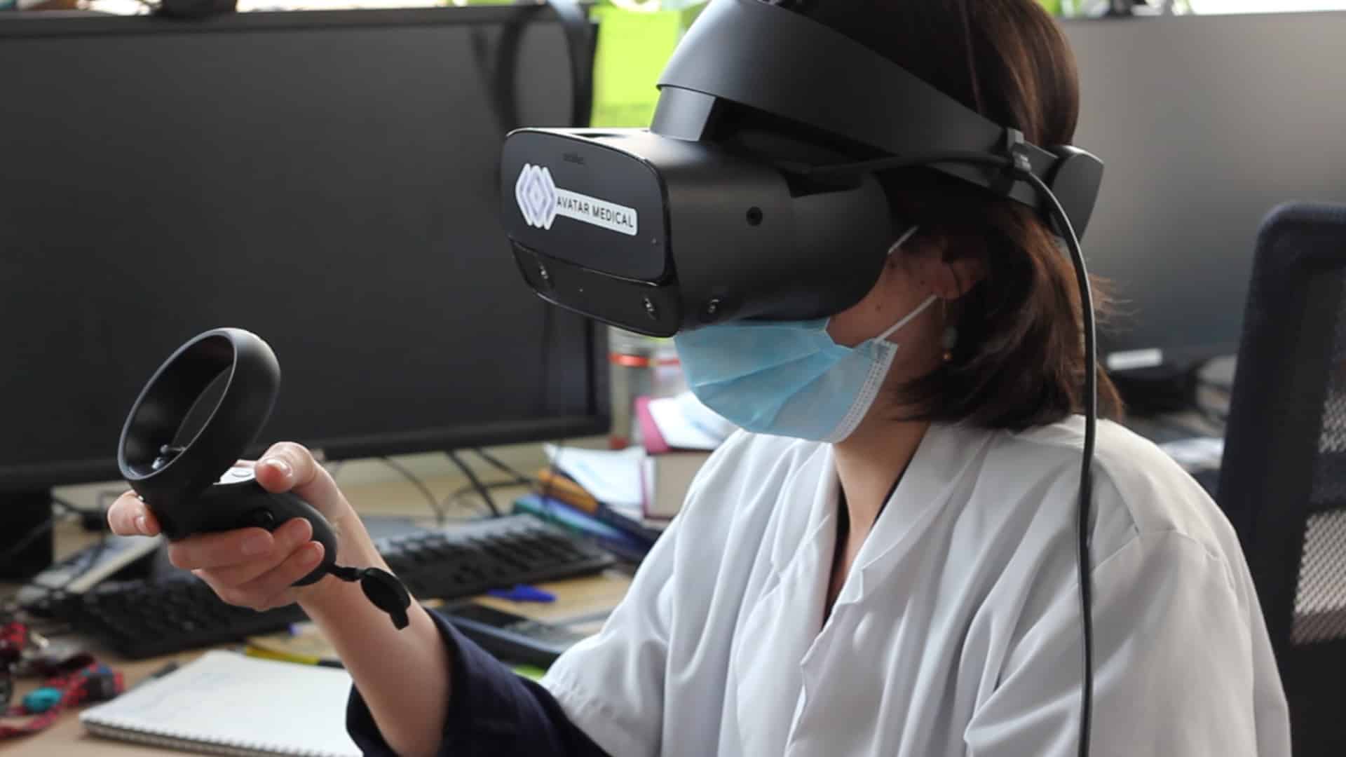
AVATAR MEDICAL‘s software transforms these files into three-dimensional representations, using a virtual reality headset. It is intended to be used before operating on patients suffering from breast cancer. Doctors and surgeons can now view 3D breast MRIs. They can navigate through them, get closer, observe the tumor from all sides, using smart filters for increased accuracy, and thus decide on the most appropriate surgical strategy.
A study conducted with 9 interns and 9 surgeons from the Institut Curie showed that AVATAR MEDICAL‘s software reduced interpretation errors fourfold and increased time savings by the same proportion.
“Today, surgical teams study 3D CT and MRI scans with sections and renderings on 2D screens. This is far from optimal for our human brain. AVATAR MEDICAL‘s software offers the surgeon user-friendliness and speed for preoperative planning, and increases productivity,” says Elodie Brient-Litzler, COO of AVATAR MEDICAL.
The software also forms part of a therapeutic de-escalation strategy. For example, it was recently used to support a partial mastectomy decision for a clinical case at the Institut Curie, thus avoiding overly invasive surgery.
This solution also applies to research and teaching and speeds up the development of young doctors’ skills.
These avatars can be created from any 3D image file. The indication can therefore be extended to other types of cancers and pathologies. In 2020, this new technology was tested in pilot sites in France and the United States for cardiovascular applications as well as in shoulder and sarcoma surgery.
The software is expected to receive marketing approval in late 2022 in the United States and early 2023 in Europe.


